Summer Research: Stories from the Microscope
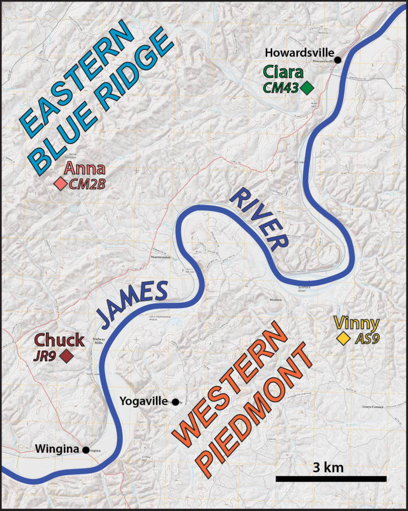
The Buckmarlson Banshees have spent much of the past seven weeks in the field working to understand the geology of the eastern Blue Ridge and western Piedmont. But this past week we came indoors and spent time observing our samples under the petrographic microscope. Earlier in the summer we cut rock samples into small chips that were then glued to a glass slide and ultimately sawed and ground down to a thickness of ~30 um (that is a third of a millimeter or one-hundredth of an inch). The final products of this process are thin sections and under the microscope both the mineralogy and texture of a sample are beautifully revealed. What follows are some geologic short stories compiled from our microscopic work.
Ciara Mills ’15 (W&M Geology) – ♫Baby I can see your halo…♫
Just west of Howardsville, things got heated up. Some 550 million years ago, much of central Virginia was covered with thousands of square kilometers of basaltic lava caused by rifting of an ancient supercontinent. These ancient lavas, now called the Catoctin Formation, look quite different than when they formed: instead of being a dark fine-grained rock, they are now many shades of green thanks to regional metamorphism.
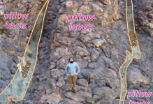
However, some of these old metabasalts still retain their original volcanic textures. Sample CM43 comes from an outcrop along the Rockfish River at the eastern edge of the Catoctin volcanic province. The hyaloclastic texture and microstructures in this sample indicates that the lava was extruded in a subaqueous environment, likely at the bottom of the Iapetus Ocean. At the outcrop scale we commonly see relict pillow structures and hyaloclastic textures develop as sea water rapidly quenches glassy rind on the pillows producing shattered and broken shards.
In this thin section, a mass of chlorite is rimmed by an irregular halo of epidote. This strange array of minerals is actually the ghost of a vesicle, or gas bubble, that formed during the cooling of the lava. This structure is now an amygdule, an original vesicle that was filled with secondary minerals (in this case, chlorite and epidote) during metamorphism and deformation. The epidote has a high birefringence, meaning that it displays vivid colors when exposed to cross-polarized light (and bears a stunning resemblance to stained glass!).
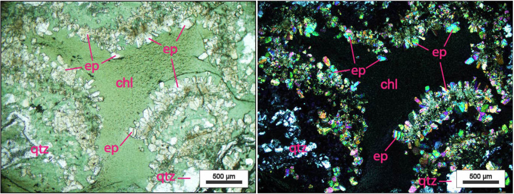
Plane-polarized (left) and crossed-polarized (right) view of amygdule in hyaloclastic greenstone from the Catoctin Formation, eastern Blue Ridge, Virginia. This amygdule contains chlorite (chl) and epidote (ep) with some quartz grains (qtz) scattered throughout.
I suspect Beyoncé wasn’t referring to epidote haloes in her song, but hey, birefringent minerals that recrystallized inside a Neoproterozoic gas bubble can be just as cool, or perhaps even cooler, than Jay-Z!
Vincent Cunningham ’16 (W&M Geology) – More Than Meets The Eye
When the Buckmarlson Banshees aren’t criticizing my campsite cooking abilities, we spend our days mapping what seems to be a sea of phyllites in the western Piedmont. Phyllites are fine-grained foliated metamorphic rocks that were originally muddy sedimentary rocks. Given the fine-grained nature of these rocks understanding their structure in the field, in the middle of bear-dwelling and tick-infested woods, is often difficult.
Sample AS9 is a phyllite that, in hand sample, appears to be composed of muscovite-rich and fine-grained quartz layers. However, when viewed more closely under the petrographic microscope, tiny eye-shaped porphyroclasts reveal all. Porphyroclasts are older mineral grains preserved in a metamorphic rock that are larger and stronger than the surrounding matrix.
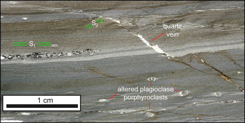
Thin section view of phyllonite from the Howardsville area, western Piedmont, Virginia. Note: porphyroclasts and multiple deformation fabrics.
As this rock stretched during deformation, the greater strength of the porphyroclasts (in this instance altered plagioclase feldspar) allowed them to maintain their shape, while the weak muscovite-rich matrix flowed around it. As a result, the porphyroclast rotated while newly precipitated minerals created “tails” parallel to the direction of stretching.
A secondary cleavage (S2) oriented at a steep angle to the primary foliation (S1) is also visible as is a younger quartz vein that offsets the earlier formed foliation. This high degree of ductile deformation transformed the phyllite into a phyllonite. Phyllonites are associated with high-strain zones, or areas of intense deformation and faulting. Studying these rocks may lead to new discoveries about the major tectonic boundaries in the western Piedmont.
Even if they won’t let me cook dinner, it’s safe to say that all the Banshees agree: phyllite eyes never lie.
Anna Spears ’15 (W&M Geology) – Staring at a Chloritoid Void
Much like the rock in Vincent’s thin section, CM2B is another phyllitic rock from the Howardsville quadrangle. If you look closely at this sample from Horse Mountain, you’ll see a well-foliated rock, but also scads of tiny eyes peering out at the geologist’s soul. Kind of creepy! Just what are these curious eyes?
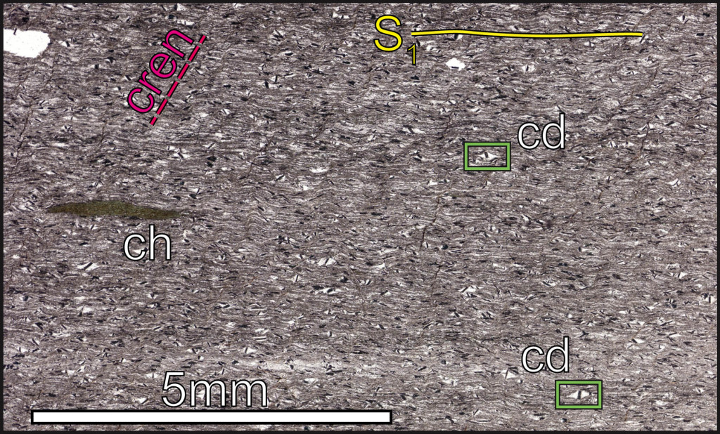
Thin section view of chloritoid (cd) ‘eyes’ in a phyllite. The sample also contains chlorite (ch), the main foliation is S1 and is cut by a crenulation cleavage (cren).
These eye-like structures are chloritoid porphyroblasts. Chloritoid is a metamorphic mineral, a hydrous Fe-Mg bearing alumino-silicate. It was originally named because of its similar appearance to chlorite, but chloritoid is a much harder mineral, topping out at 6.5 on Mohs hardness scale. Porphyroblasts are larger mineral grains that have grown during metamorphism and deformation (as opposed to porphyroclasts which are older than the matrix). Phyllites typically are not erosionally resistant rocks, but these chloritoid-bearing phyllites underlie a distinct northeast/southwest trending ridge in the eastern Blue Ridge.
CM2B’s story begins long ago (likely during the Cambrian period ~500 Ma) as mud that accumulated off the continental margin of ancient North America. By the mid-Paleozoic (350 to 320 Ma) the rock had been buried to sufficient depths that the clay minerals recrystallized to form muscovite and chlorite. Additionally, there must have been enough iron to facilitate the growth of chloritoid porphyroblasts. These new metamorphic minerals grew while the rock was deforming which resulted the main foliation.
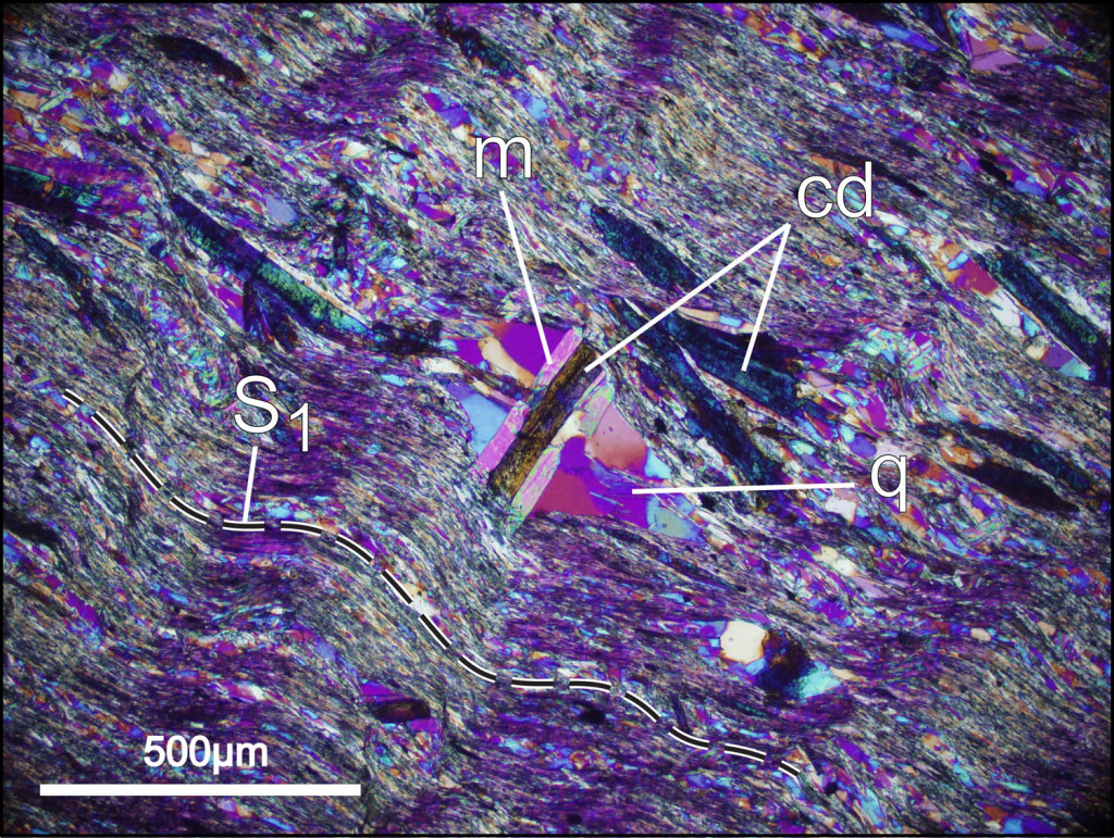
Close-up view in cross-polarized light with the gypsum plate inserted. cd- chloritoid, m- muscovite, q- quartz, S1 is the main foliation
But the story doesn’t end there. This phyllite was deformed AGAIN! A later deformation episode resulted in the rotation of chloritoid grains and the development of pressure shadows. To complicate things further, the earlier-formed foliation was also crinkled and crenulated.
Chuck Bailey ’89 (W&M Geology) – Up Close with a Red Bed
As the Buckmarlson Banshees have noted the eastern Blue Ridge and western Piedmont is awash in metamorphic rocks, but two areas underlain by Mesozoic sedimentary rocks in the Scottsville and Midway Mills basins occur within the study area. These are among the youngest rocks in central Virginia.
Sample JR9 is a fine-grained sandstone with a silty matrix. At the outcrop scale these Mesozoic (likely Triassic) sandstones and siltstones are distinctly reddish and in the geological vernacular known as red beds. At the scale of a thin-section (~40 mm by 20 mm) the ruddy nature of the matrix is striking. The red material includes hematite, an iron-oxide mineral that forms as the result of weathering and oxidation of Fe-bearing minerals, and makes the world’s red beds – red. The question is did hematite form as a primary mineral when the sediment was deposited or was it precipitated at a later date by ground waters carrying Fe in solution?
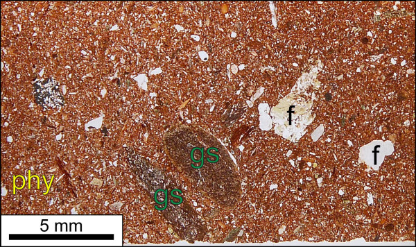
Thin section view of red silty sandstone from the Midway Mills Mesozoic basin, central Virginia. Note clasts of feldspar (f), greenstone (gs), and phyllite (phy).
The clasts also tell a story, they include angular fragments of feldspar and quartz, some rounded pieces of greenstone, and even a few chips of phyllite. The source area for these Mesozoic basins was the surrounding metamorphic rocks of the eastern Blue Ridge and western Piedmont. The angular and poorly sorted nature of this detritus indicates that sediment was transported only short distances prior to being deposited.
Comments are currently closed. Comments are closed on all posts older than one year, and for those in our archive.

Looks like there are several cool new projects going on in the Structure&Tectonics lab! Good luck to the Buckmarlson Banshees (not that they’ll need it!)